


2023.04.24
Report: The 13th BRI International Symposium
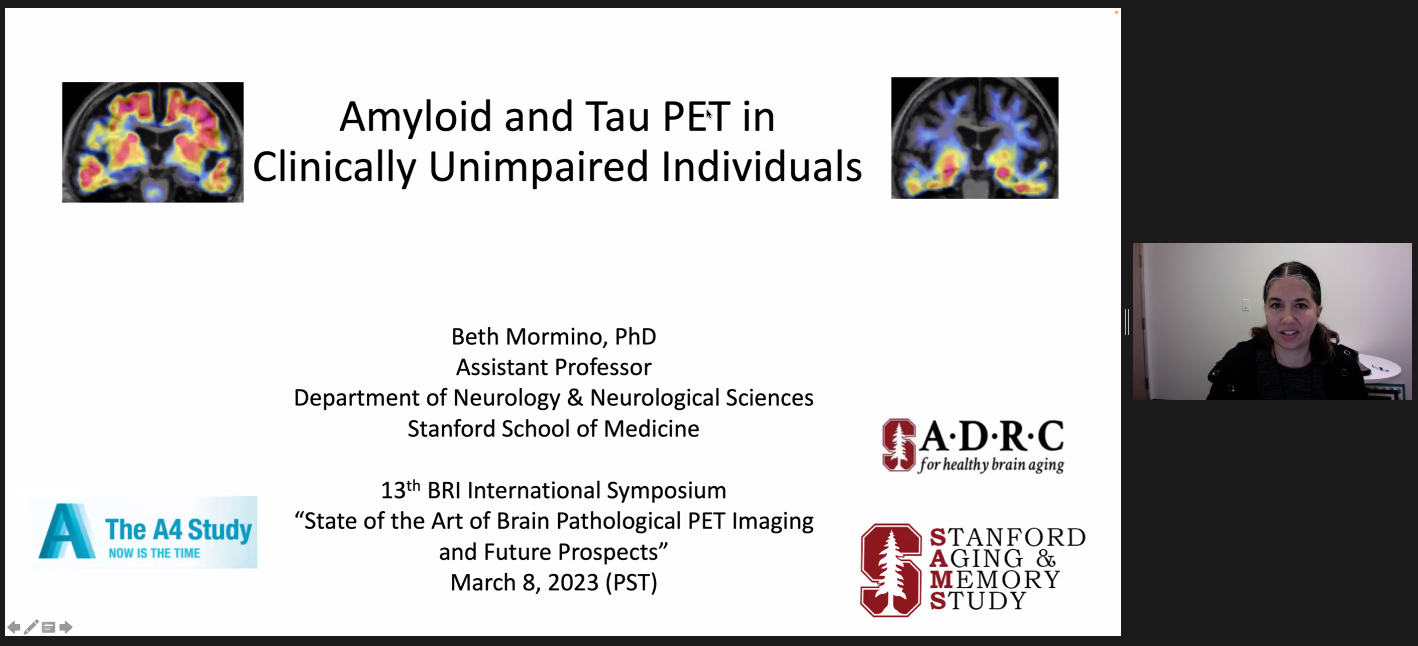
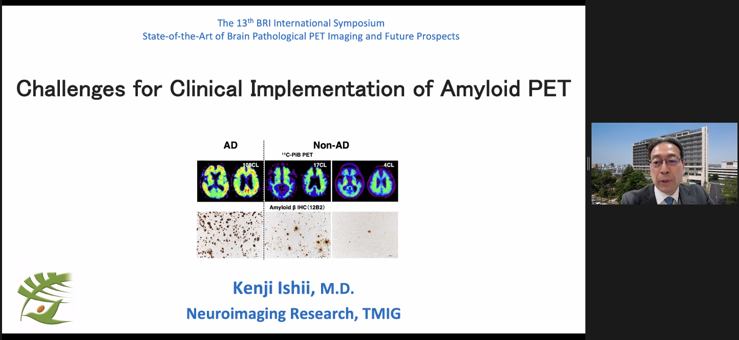
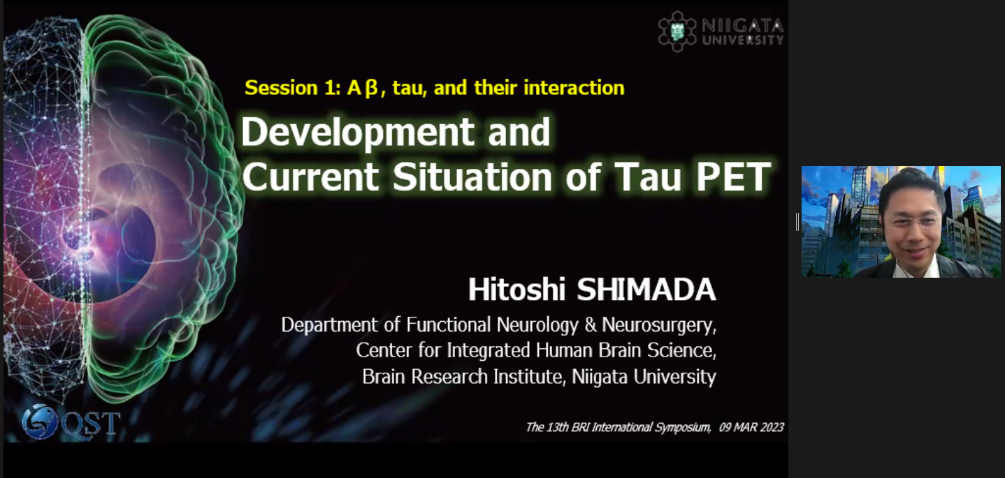
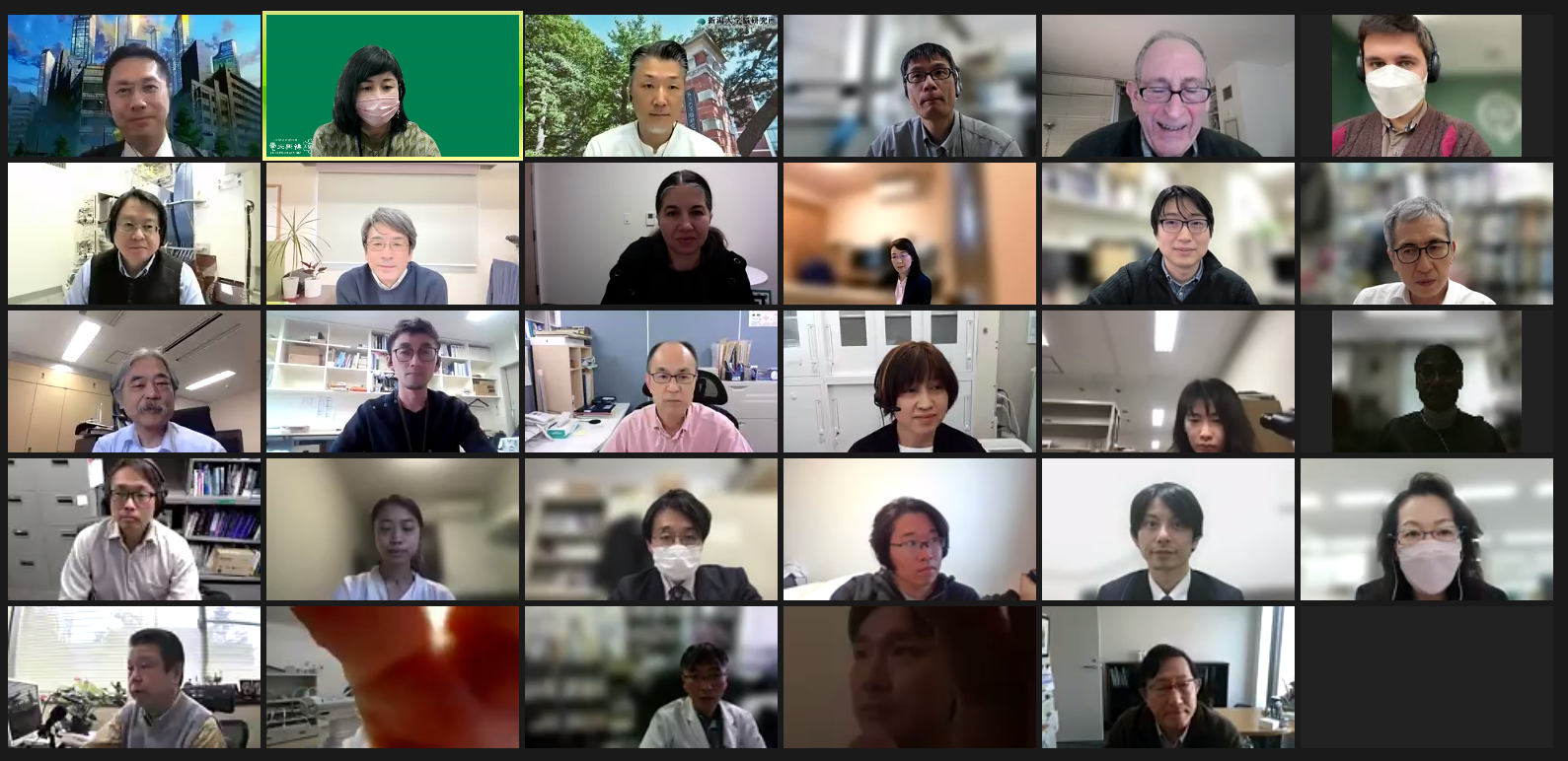
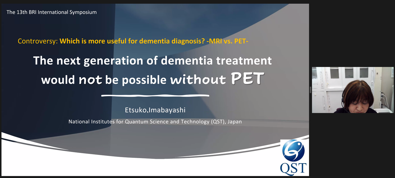
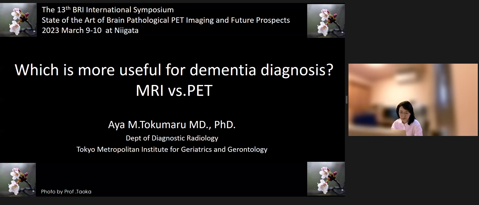
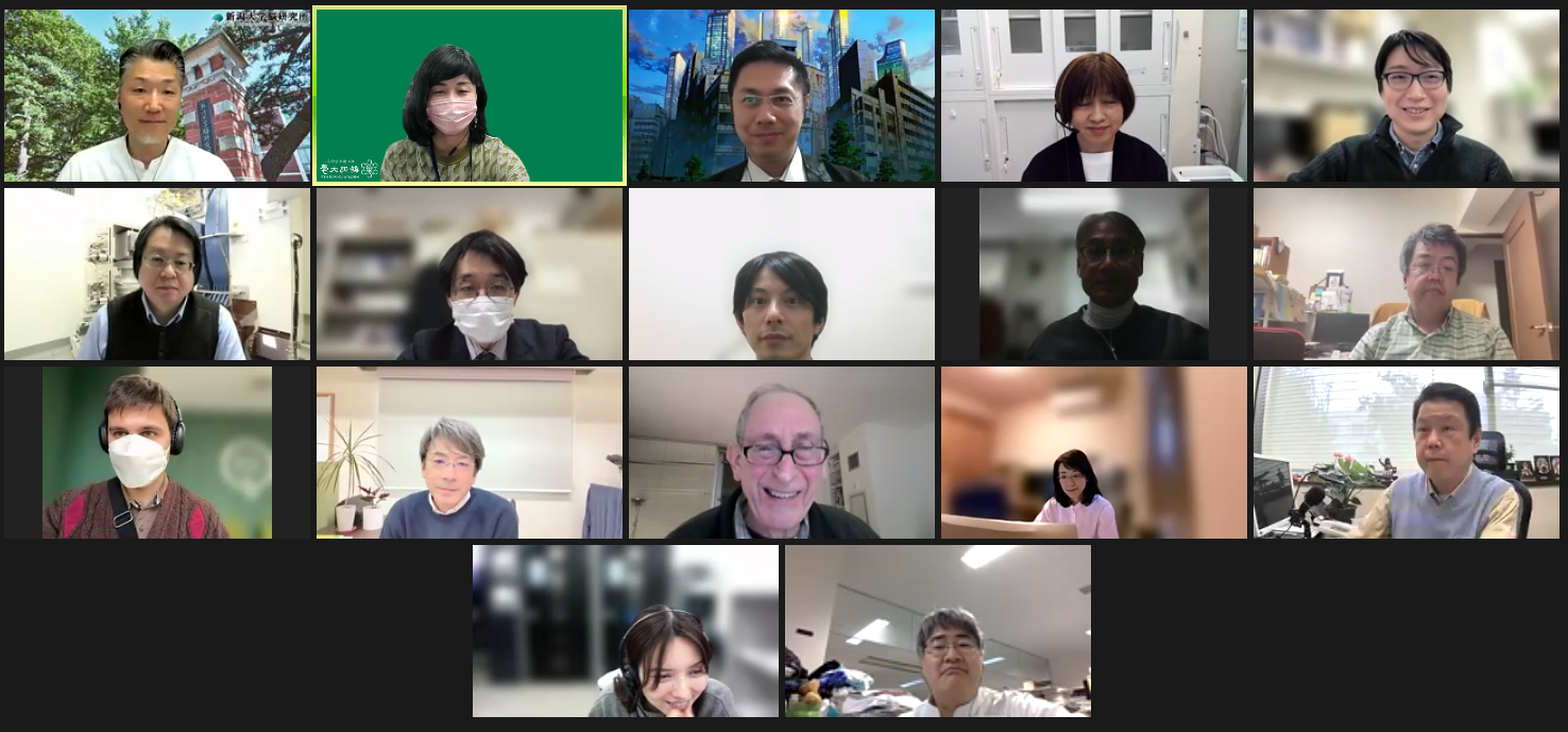
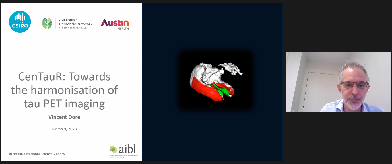
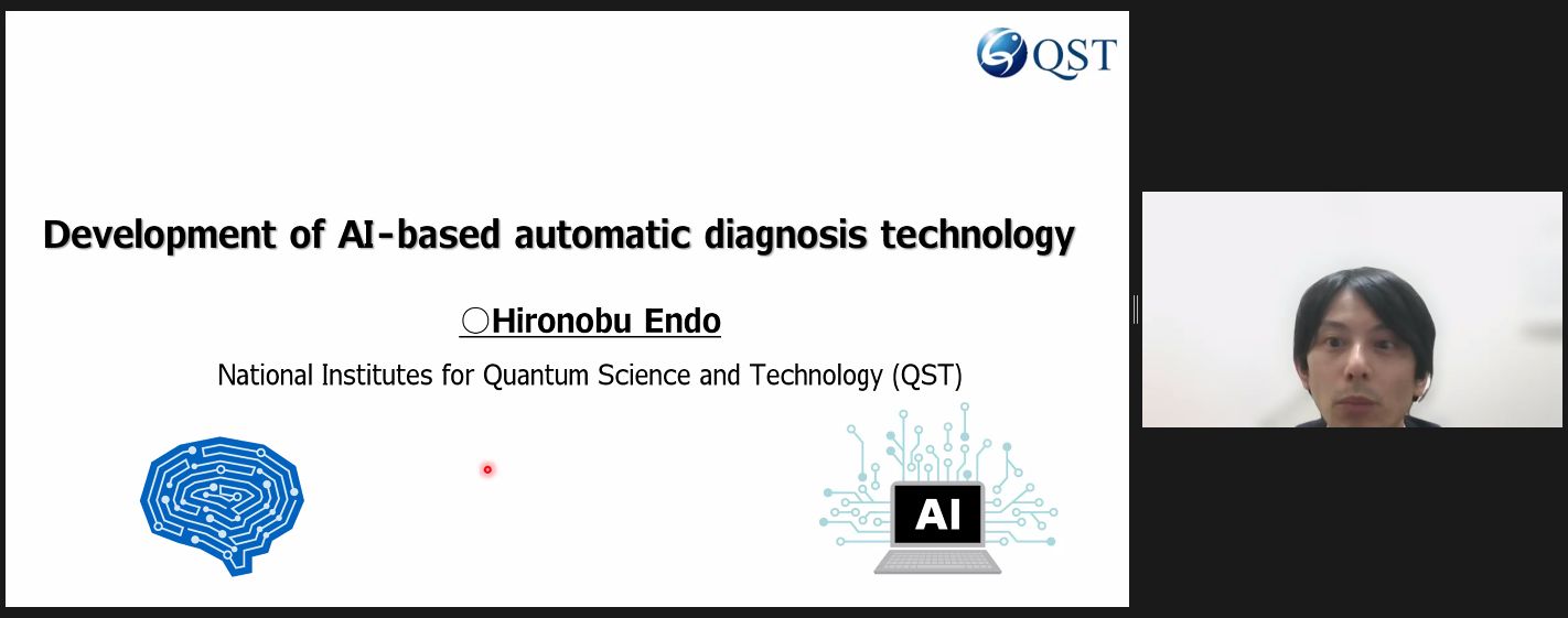
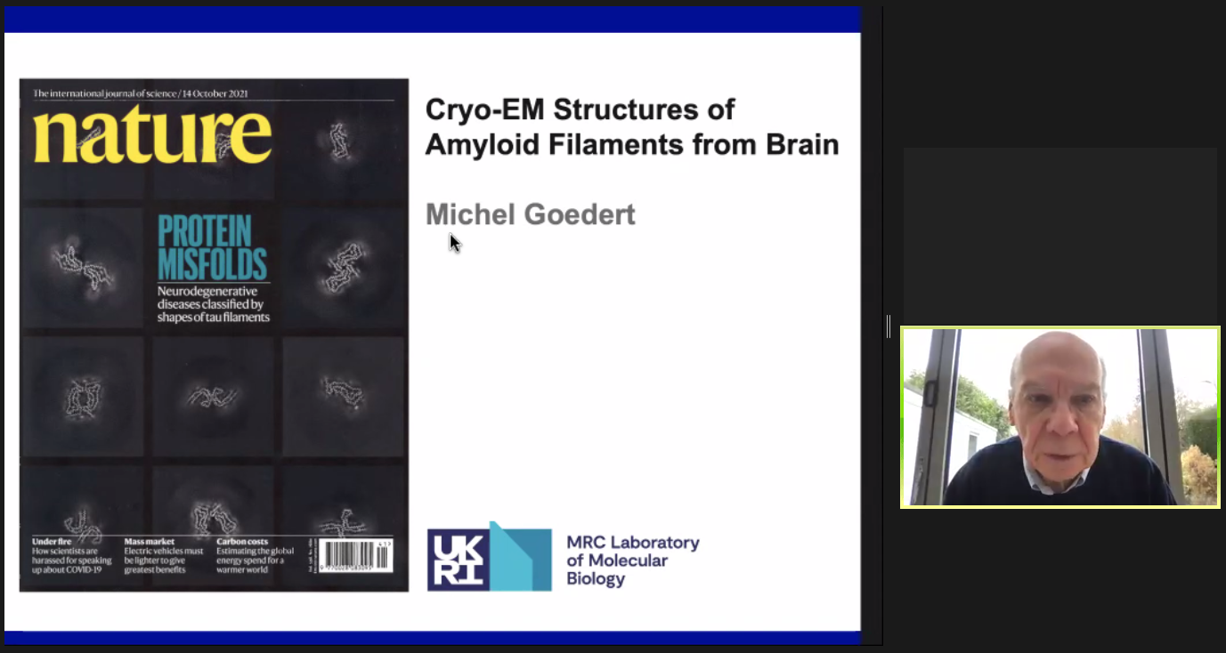
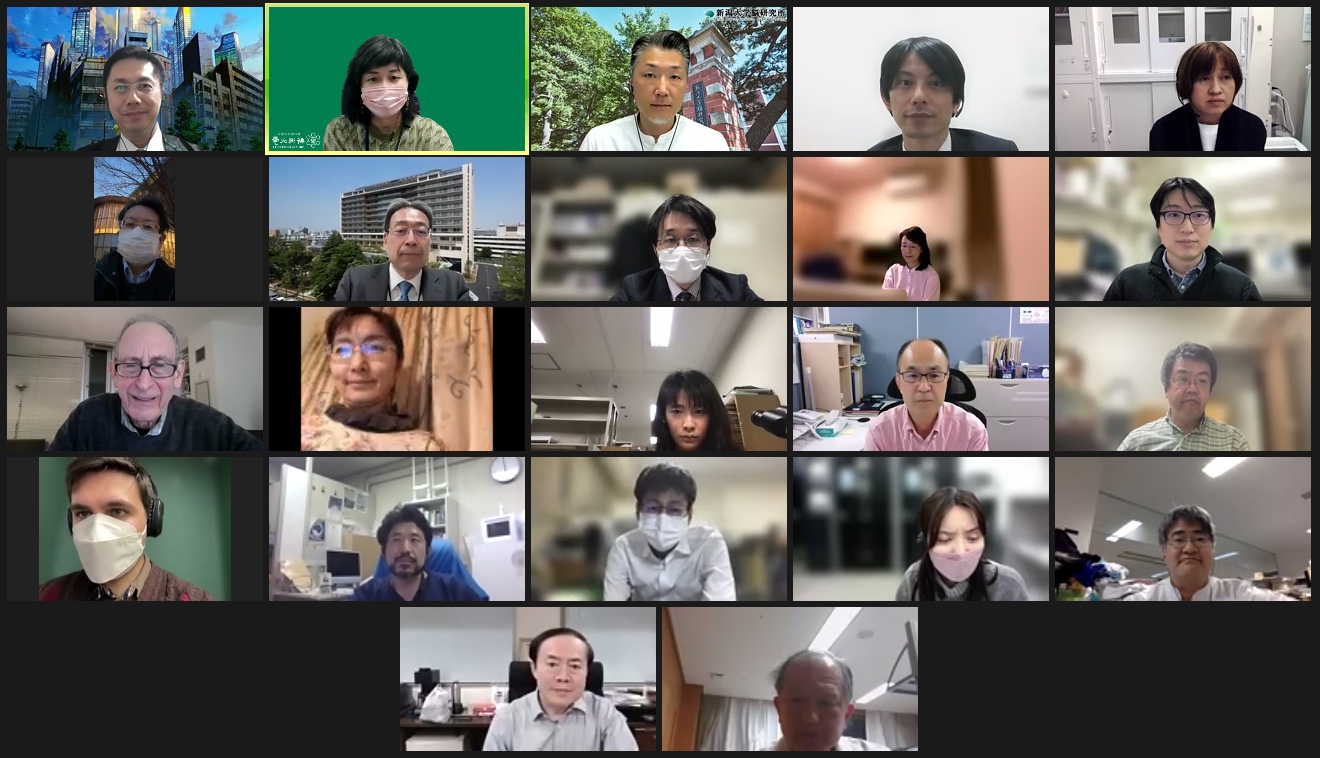
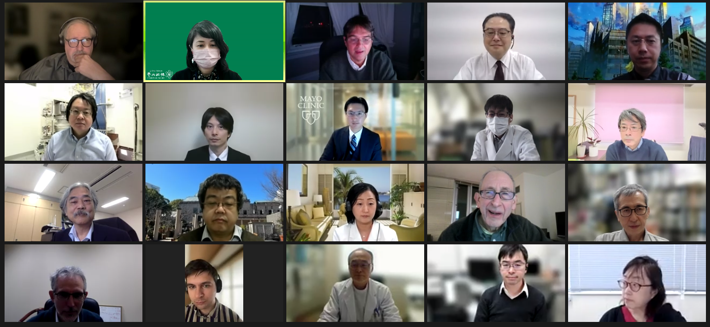
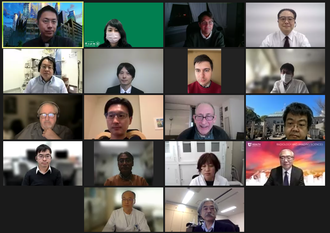
The 13th annual BRI international symposium was held online on March 9-10, 2023. The topic of this symposium was "State-of-the-Art of Brain Pathological PET Imaging and Future Prospects." There were eight speakers from abroad - Dr Elizabeth Mormino (Stanford University, US), Dr Vincent Doré (Commonwealth Scientific and Industrial Research Organisation (CSIRO), Australia), Dr Michel Goedert (MRC Laboratory of Molecular Biology, UK), Dr Michael J. Pontecorvo (Avid Radiopharmaceuticals, US), Dr Victor Villmagne (University of Pittsburgh, US), Dr Shunsuke Koga (Mayo clinic, US), Dr Pedro Rosa-Neto (McGill University, Canada), Dr Satoshi Minoshima (University of Utah, US) , and seven speakers from Japan - Dr Kenji Ishii (Tokyo Metropolitan Institute of Gerontology (TMIG)), myself, Dr Etsuko Imabayashi (National Institutes for Quantum Science and Technology (QST)), Dr Aya M. Tokumaru (TMIG), Dr Hironobu Endo (QST), Dr Nobuyuki Okamura (Tohoku Medical and Pharmaceutical University), and Dr Maiko Ono (QST). To our great joy, a total of 233 people joined the two-day symposium (75% from outside Niigata University, including overseas participants from four countries).
In recent years, brain pathology imaging technology has played a pivotal role in elucidating the pathology of Alzheimer's disease and other neurodegenerative disorders by visualizing abnormal proteins such as amyloid-beta and tau aggregates as well as neuroinflammation. This symposium pays homage to the Human Amyloid Imaging Conference (HAI), the world's largest international conference, which has contributed to the acceleration of research in this field over the past 15 years, and we planned and prepared for the symposium, hoping to provide the same high-level lectures and lively discussions that we have seen at HAI.
The four thematic sessions covered:
(1) the latest topics related to PET imaging of amyloid-beta and tau protein lesions, the two major pathological changes in Alzheimer's disease;
(2) the development of methods for comparing different tau PET ligands, automated diagnostic methods using AI, and 3D structural identification of pathological proteins using cryo-EM;
(3) Challenges and pitfalls in clinical implementation of amyloid PET and tau PET; and
(4) Novel imaging techniques targeting epigenomic alterations, astroglia activity, alpha-synuclein aggregates, among others.
The top experts gave presentations and engaged in lively discussions. During the controversy session, the discussion on the usefulness of diagnostic imaging techniques in the treatment of dementia by speakers from the standpoints of MRI and PET was a new challenge for this symposium, and we believe that this session gave us a sense of the various possibilities that lie ahead for future symposia. The symposium concluded with a special lecture by Dr Satoshi Minoshima, who provided a valuable opportunity for the audience to learn about the past and present state of brain imaging analysis and where we stand today
We would like to thank all the speakers, attendees, and administrative staff members for their great contributions to making the symposium successful.
By Prof Hitoshi Shimada
(Dept of Functional Neurology & Neurosurgery)
| click to enlarge image | ||||
 |
 |
 |
 |
 |
 |
 |
 |
 |
 |
 |
 |
 |
 |
 |
 |
 |
 |
 |
 |
