


Members
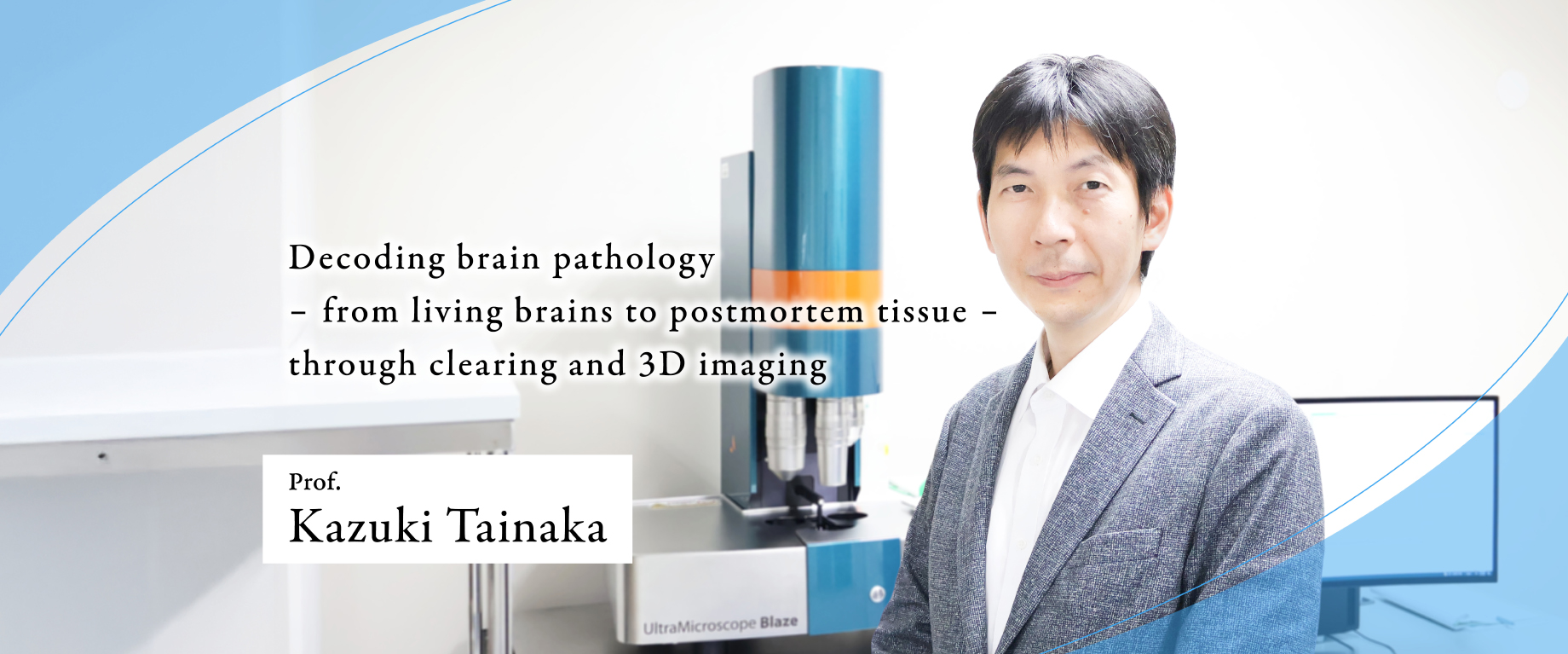
Kazuki TAINAKA
Hitoshi UCHIDA
Xinyi LIU
Research Focus
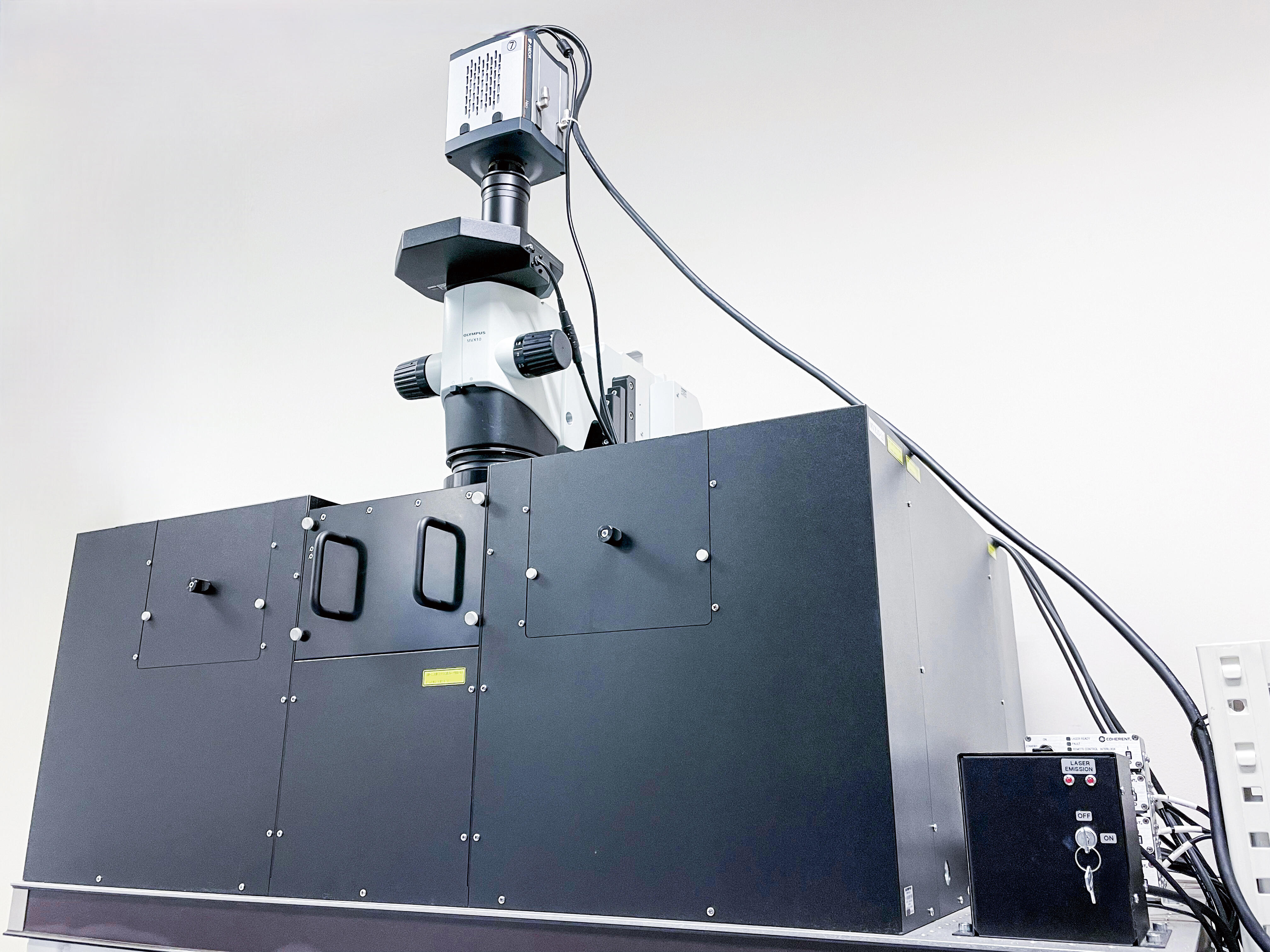
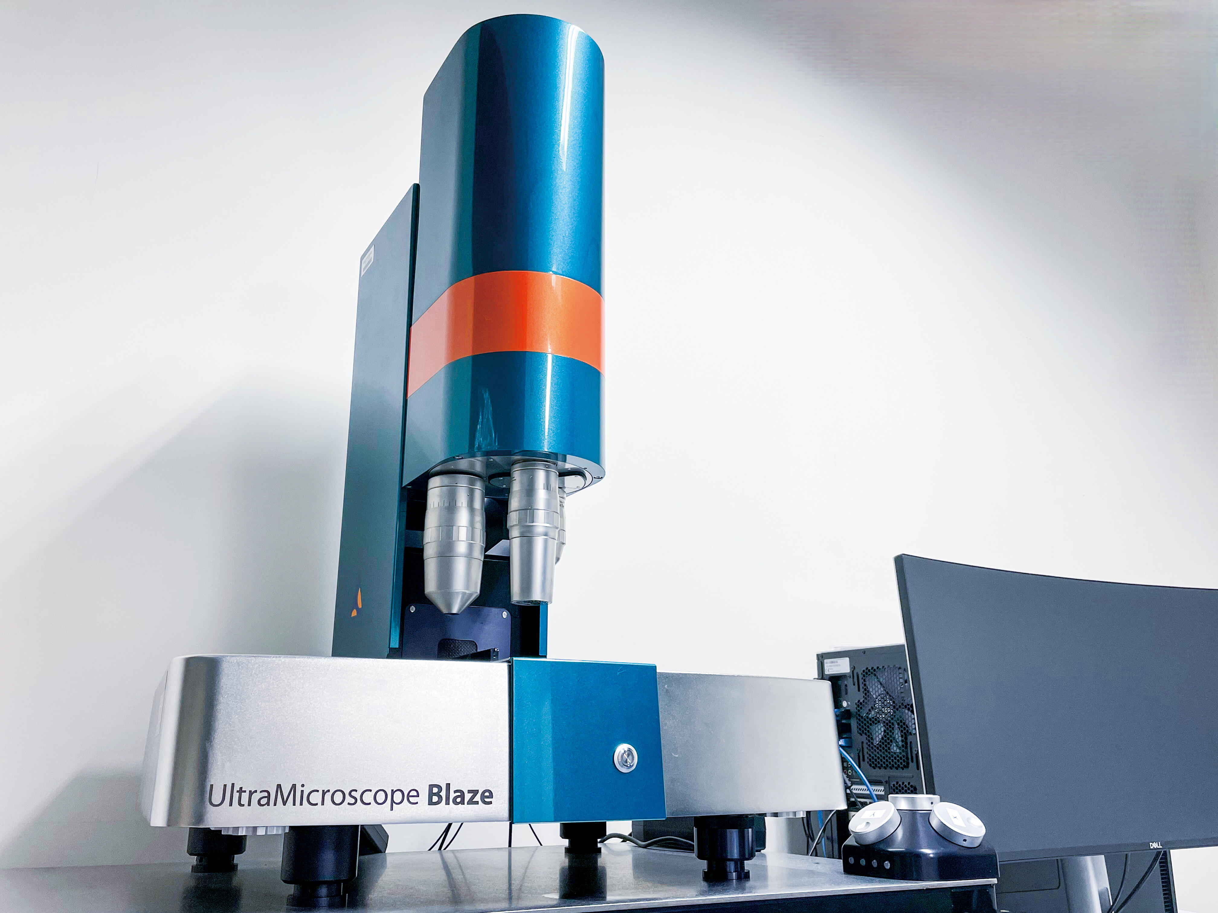
Our department develops tissue clearing techniques combined with high-speed three-dimensional fluorescence imaging to establish "3D neuropathology" of the brain and other tissues. Recently, we developed SeeThrough, a skull-clearing reagent that enables minimally invasive, longitudinal imaging of the living mouse brain without skull removal (Liu et al., Nat Commun, 2025). We also rationally designed an activatable covalent fluorescent probe compatible with tissue clearing, enabling whole-body and whole-organ 3D mapping of tissue hypoxia (Sakamoto et al., ACS Nano, 2024). In an international collaboration, we have further contributed to whole-brain cell mapping by combining highly multiplexed in situ hybridization with AI-driven analysis (Murakami et al., Neuron, 2026). Building on these tissue-clearing platforms, we aim to elucidate disease mechanisms and translate these technologies into next-generation diagnosis and treatment of neurological disorders.