


2026.02.10
From surface to depth: 3D imaging traces vascular amyloid spread in the human brain
Niigata, Japan - Researchers at Niigata University have used advanced three-dimensional (3D) imaging to reveal how amyloid β (Aβ) deposits spread along blood vessels in the human brain in cerebral amyloid angiopathy (CAA). By analyzing postmortem brain tissue, the team showed that vascular amyloid deposition is most prominent in surface arteries and extends continuously toward deeper vascular branches, providing insight into the spatial organization of amyloid pathology in CAA.
CAA is a cerebrovascular disorder characterized by Aβ deposition in cerebral small vessel walls. It is a major cause of lobar intracerebral hemorrhage and frequently coexists with Alzheimer's disease. Although impaired clearance of Aβ along vascular pathways is thought to contribute to CAA, how vascular amyloid deposition is spatially distributed across the brain's vascular network has remained poorly understood.
To address this question, researchers analyzed postmortem brain tissue from six patients with CAA using tissue-clearing techniques and light-sheet fluorescence microscopy. This approach enabled large volumes of human brain tissue to be visualized in three dimensions while preserving the continuity of the vascular network. By fluorescently labeling vascular smooth muscle cells and Aβ, the researchers reconstructed blood vessels extending from the cortical surface to the underlying white matter.
"What makes this study unique is our ability to visualize the entire vascular network in the human brain in three dimensions and at cellular resolution," explains Dr. Saito, who led the research. "Our 3D approach revealed a continuous pattern of deposition that we couldn't have seen with conventional two-dimensional sections."
"A key finding is that amyloid doesn't deposit randomly," notes Hayashi, the study's first author. "It follows a specific pattern--starting from surface vessels and spreading inward along connected pathways. This supports the hypothesis that CAA develops due to impaired clearance mechanisms."
The researchers also found that the density of parenchymal amyloid plaques was lower around Aβ-positive vessels than in other areas, indicating spatially preferential distribution.
"Areas with heavy vascular amyloid had fewer plaques in surrounding brain tissue," Hayashi observes. "Understanding this balance could be important for developing targeted therapies."
Together, these findings provide a 3D framework for understanding how vascular amyloid pathology is organized in the human brain. The results support the idea that impaired perivascular clearance pathways contribute to the progression of CAA and highlight the importance of spatial context in amyloid-related cerebrovascular disease.
▸EurekAlert!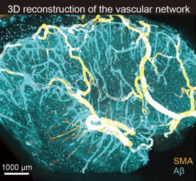
3D reconstruction of the vascular network.
Using postmortem human brain tissue, three-dimensional imaging revealed vascular Aβ deposition patterns and a lower density of parenchymal amyloid plaques surrounding Aβ-positive vessels. Smooth muscle actin (SMA) (yellow) and Aβ (blue).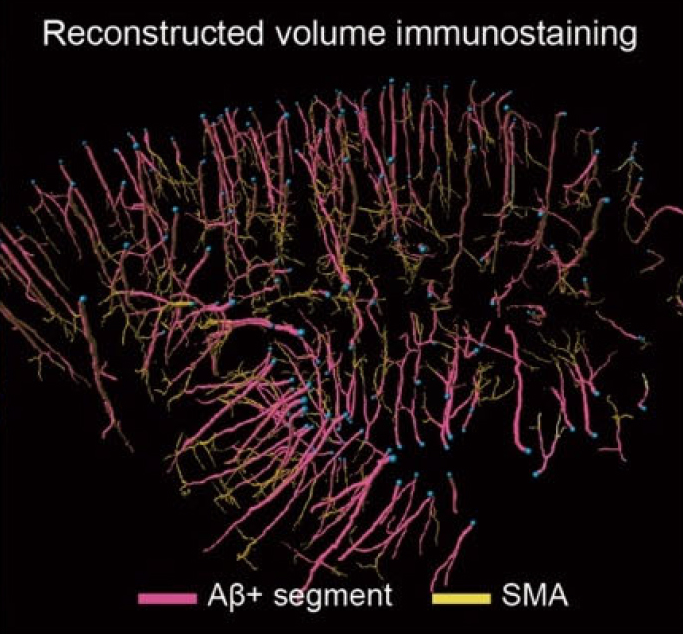
Reconstructed volume immunostaining
A schematic 3D vascular network shows Aβ-positive (pink) and Aβ-negative/SMA-positive (yellow) segments.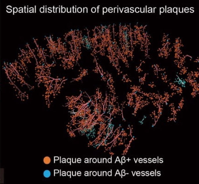
Reconstructed perivascular senile plaques around Aβ-positive/negative vessels.
Original Publication
"Expansive spatial pattern of Aβ deposition in patients with cerebral amyloid angiopathy: A three-dimensional surface-to-depth analysis"
Hayashi H, Saito R, Miyashita A, Ikeuchi T, Tada M, Akazawa K, Onodera O, Tainaka K, Kakita A.
Sci Adv. 2026 Feb 6;12(6):eaea7539. doi: 10.1126/sciadv.aea7539. Epub 2026 Feb 6.
