Development of Brain Maps and Their Disorders
The cerebellum of fish, unlike that of humans, appears uniform without
visible folds or boundaries. However, the neural circuits in the fish cerebellum
are quite similar to those in humans, with only minor differences. We have
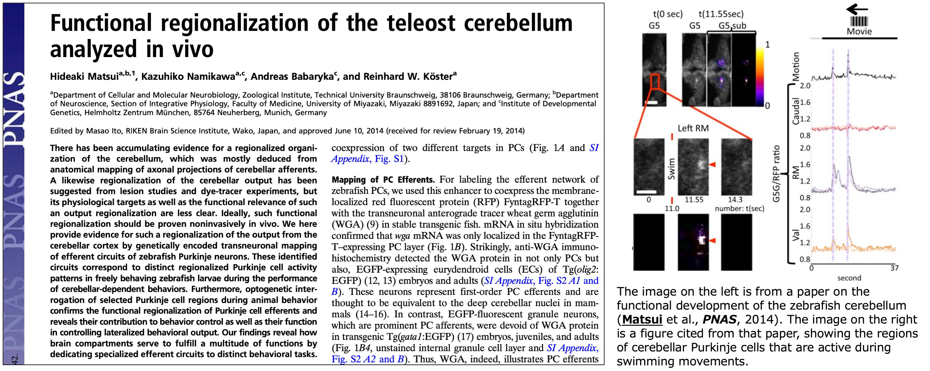
elucidated the efferent pathways of the zebrafish cerebellum using various
methods. By employing calcium imaging, we visualized neural activity specific
to swimming and nystagmus, demonstrating that the anatomical maps correspond
to maps of neural activity. Using optogenetics to intervene in neural activity
in these areas resulted in corresponding behavioral abnormalities. These
findings were published in PNAS in 2014, accompanied by editorial comments
from Dr. Hitoshi Okamoto of RIKEN (Matsuiet al., Proc. Natl. Acad. Sci. USA, 2014).
Through these efforts, we successfully mapped the functional areas of the vestibulocerebellum and spinocerebellum in fish. However, there are still unknown regions. In humans, the functional analysis of the vestibulocerebellum and spinocerebellum has progressed, but the role of the cerebellum in higher functions such as social behavior remains largely unexplored. We are currently investigating the cerebellum of fish and humans, particularly the parts involved with the cerebrum, to understand their potential deep connections with various developmental disorders. We also aims to provide deeper insights into autism and ADHD.
Autism in Fish
Autism-like alterations in social behavior are often thought to be uniquely
human. However, recent studies using animal models have revealed that their
origins and mechanisms are deeply rooted in evolutionarily conserved brain
functions and sensory processing.
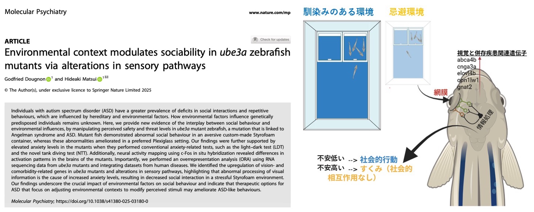
In our work, we used zebrafish lacking the autism- and Angelman syndrome–related
gene UBE3A to investigate how social behavior is shaped by environmental factors.
We compared two experimental settings: a “white polystyrene tank,” which
induces anxiety in fish, and a “transparent acrylic tank,” which resembles
a breeding environment and provides a sense of safety. We found that ube3a-mutant fish displayed reduced social interaction in the polystyrene environment
but showed improved social behavior in the acrylic environment. Gene expression
analyses and brain activity mapping further demonstrated that this reduction
in sociality was driven by abnormal visual information processing and heightened
anxiety.
These findings reveal that the social behavior of genetically predisposed
individuals can be strongly modified by environmental context. They also
suggest that in autism spectrum disorder (ASD), providing safe environments
and adjusting sensory stimuli may help improve social behaviors (Dougnon and Matsui, Molecular Psychiatry, 2025).