Facilities and model animals
Super-resolution microscopy
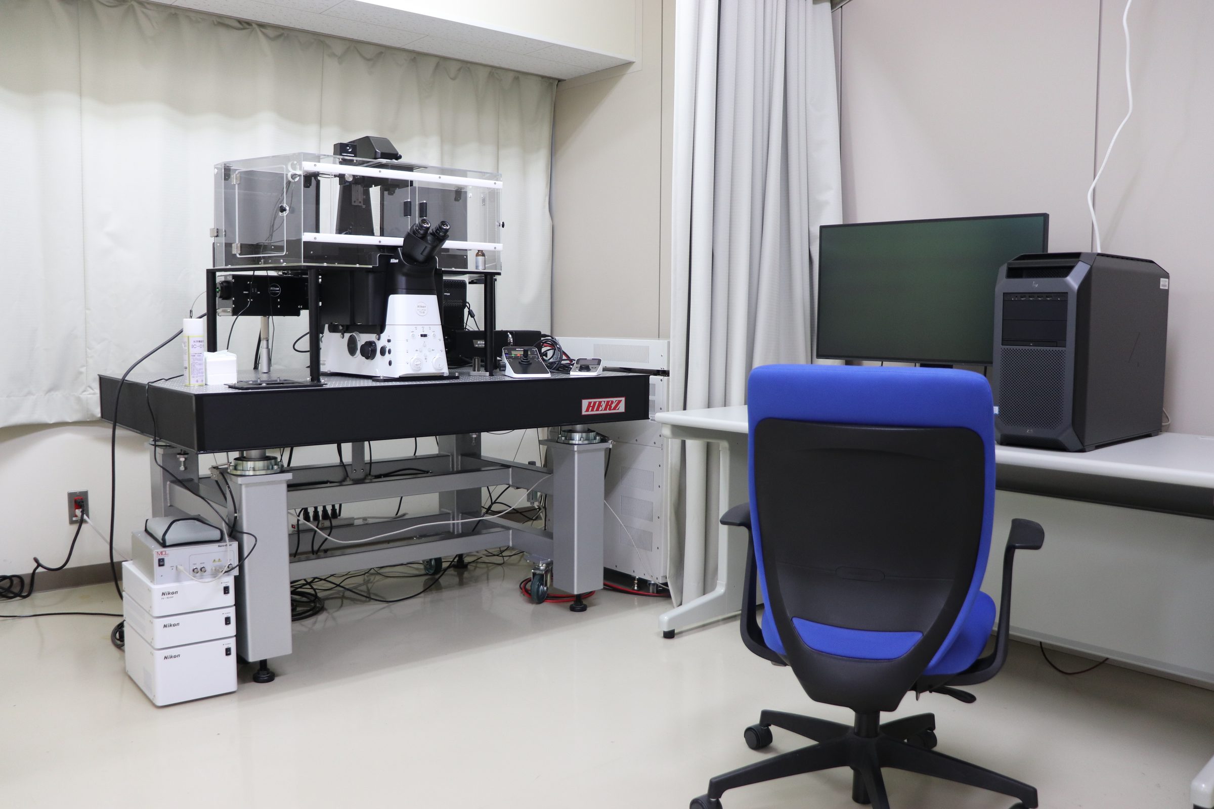
Nikon SIM + STORM. One of our higher-end instruments, part 1.
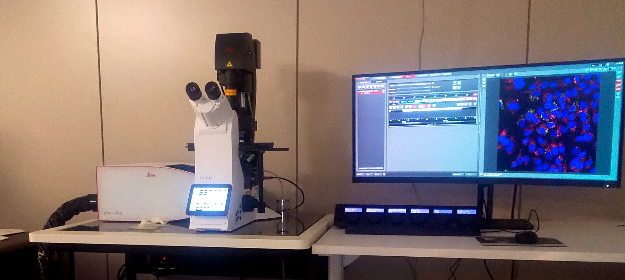
Leica STELLARIS + LIGHTNING
Computational processing makes fine structures inside cells clearer in confocal images. Multi-color fluorescence imaging is also supported.
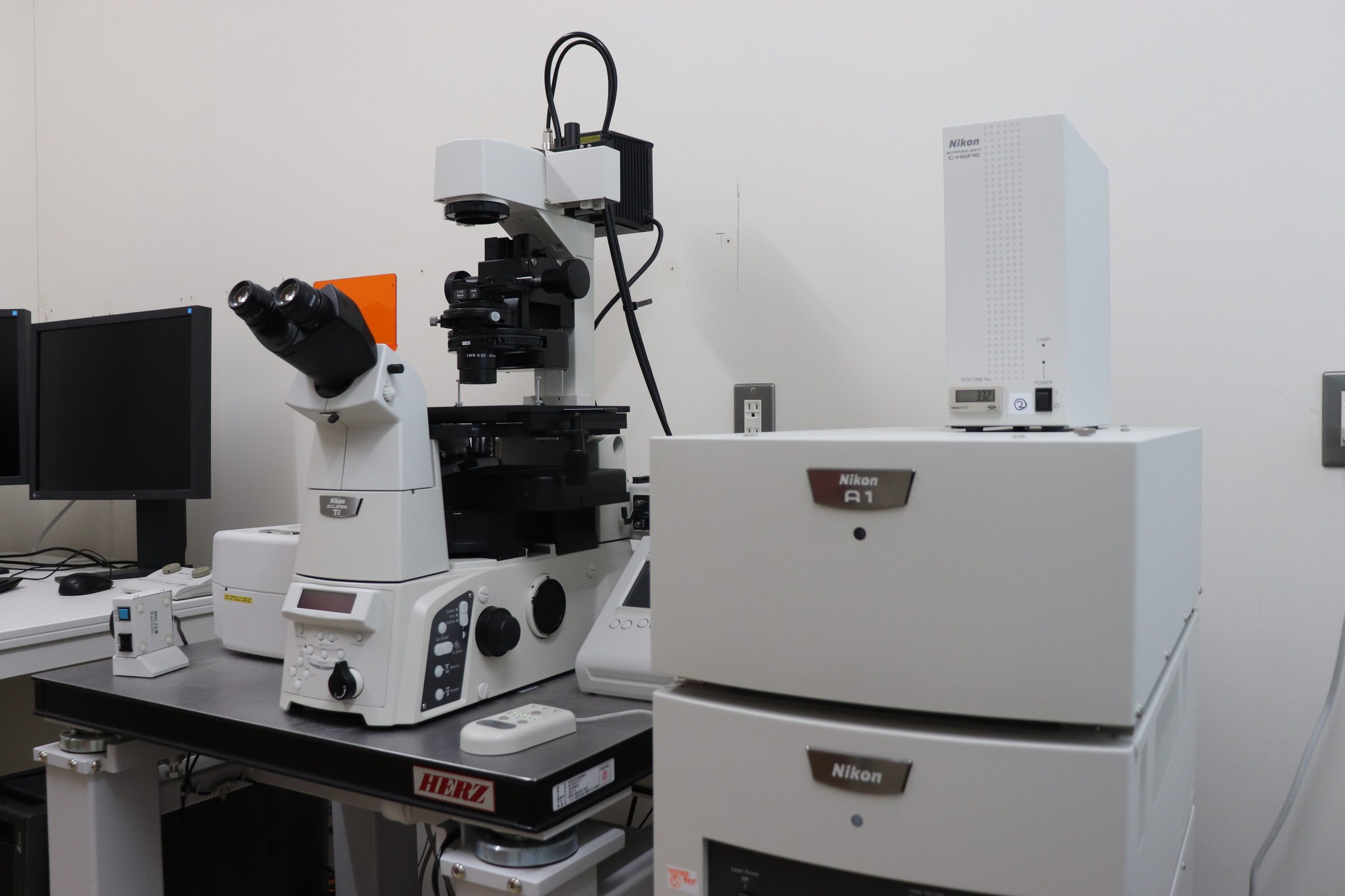
Confocal microscopy, Nikon A1R+
A fast, sensitive confocal microscope. In Matsui LAB. this is the instrument for everyday routine observation.
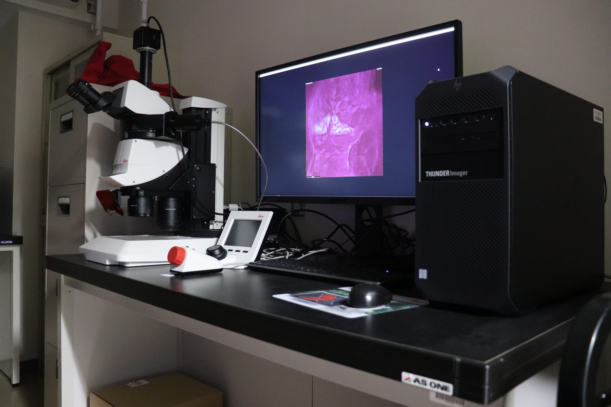
Leica THUNDER model organism
A stereo microscope that uses Computational Clearing to show fine structure clearly. It is useful when we want an overview of zebrafish or other thick samples.
High-content confocal imaging
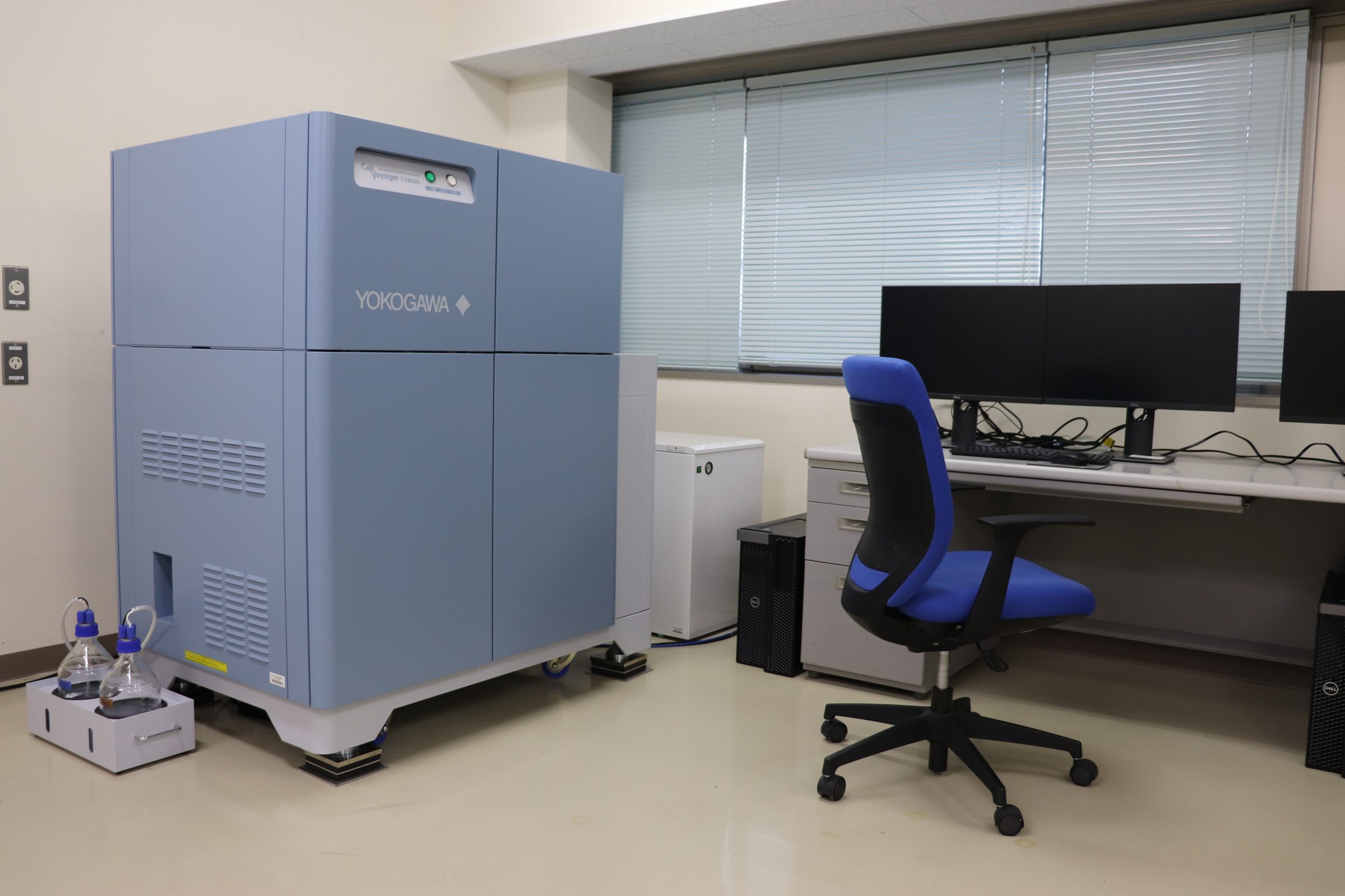
Yokogawa CV8000. Another of our higher-end instruments. It is a shared high-content imaging system managed by our laboratory.
Single Cellome Unit SU10
Yokogawa’s Single Cellome Unit SU10. A glass nanopipette with a tip tens of nanometres across delivers genes or drugs directly into the nucleus or cytoplasm of a chosen single cell, or samples material from inside it.
Basic equipment for cell culture and molecular biology
Plate readers: fluorescence, absorbance and luminescence. The lab currently has three. Nanodrop. Four PCR machines and two real-time PCR machines. Cell-culture room: two hoods, two CO2 incubators, a Nepa21 electroporator, an automatic cell counter (CellDrop).
Fish breeding system
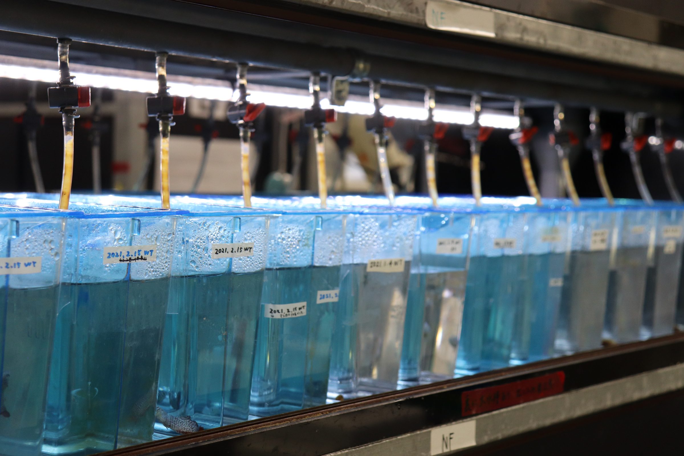
Six large double-sided four-shelf recirculating systems. We use MEITO Suien tanks.
Research models
Mouse
We use mice to test, in the mammalian brain, mechanisms found in fish and cultured cells.
Zebrafish
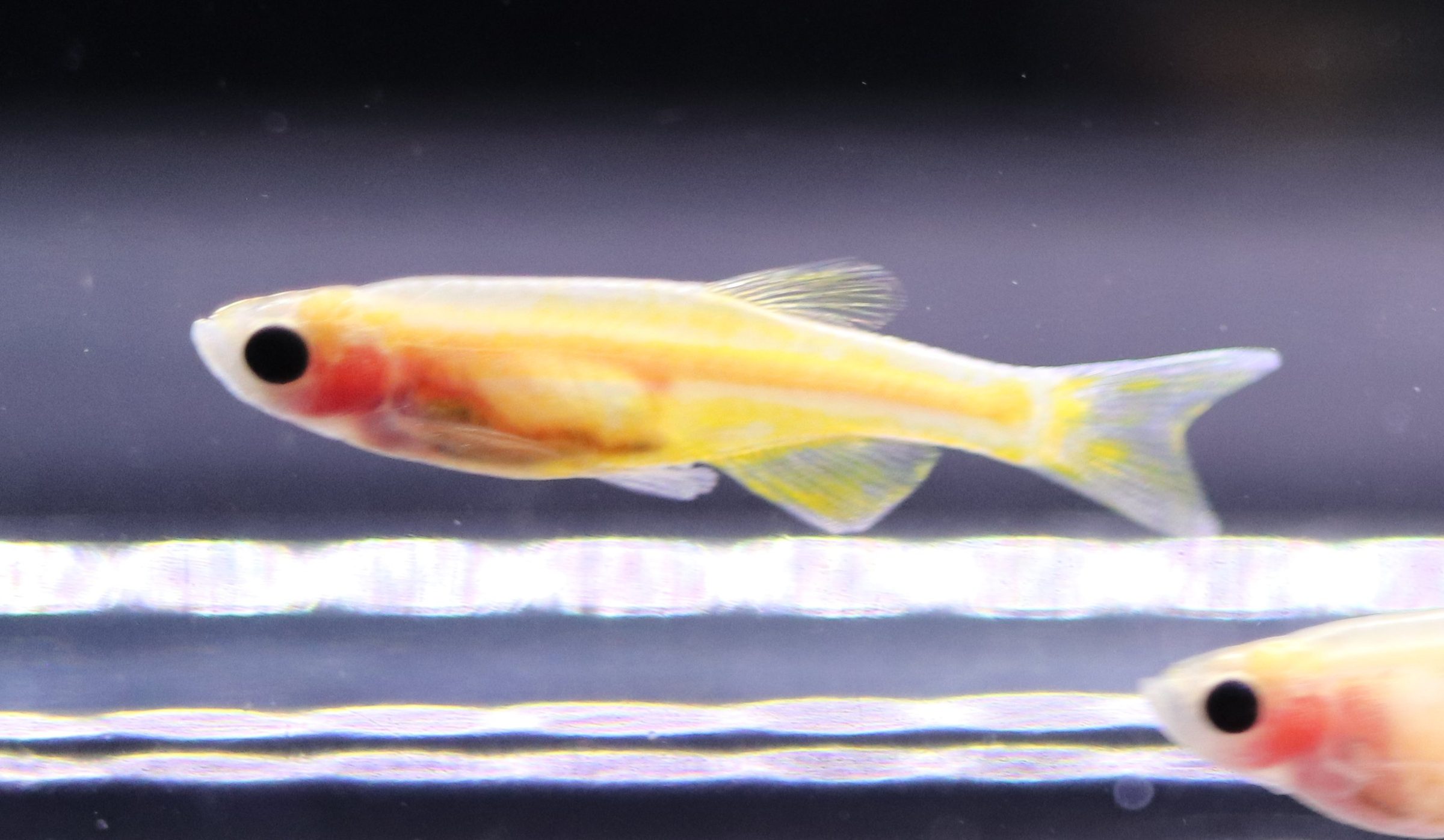
This is the fish we use most, and a vertebrate model used widely around the world. Easy husbandry and genome editing, many tools, and easy imaging make it exceptionally practical. The photograph is the transparent Casper line.
Medaka
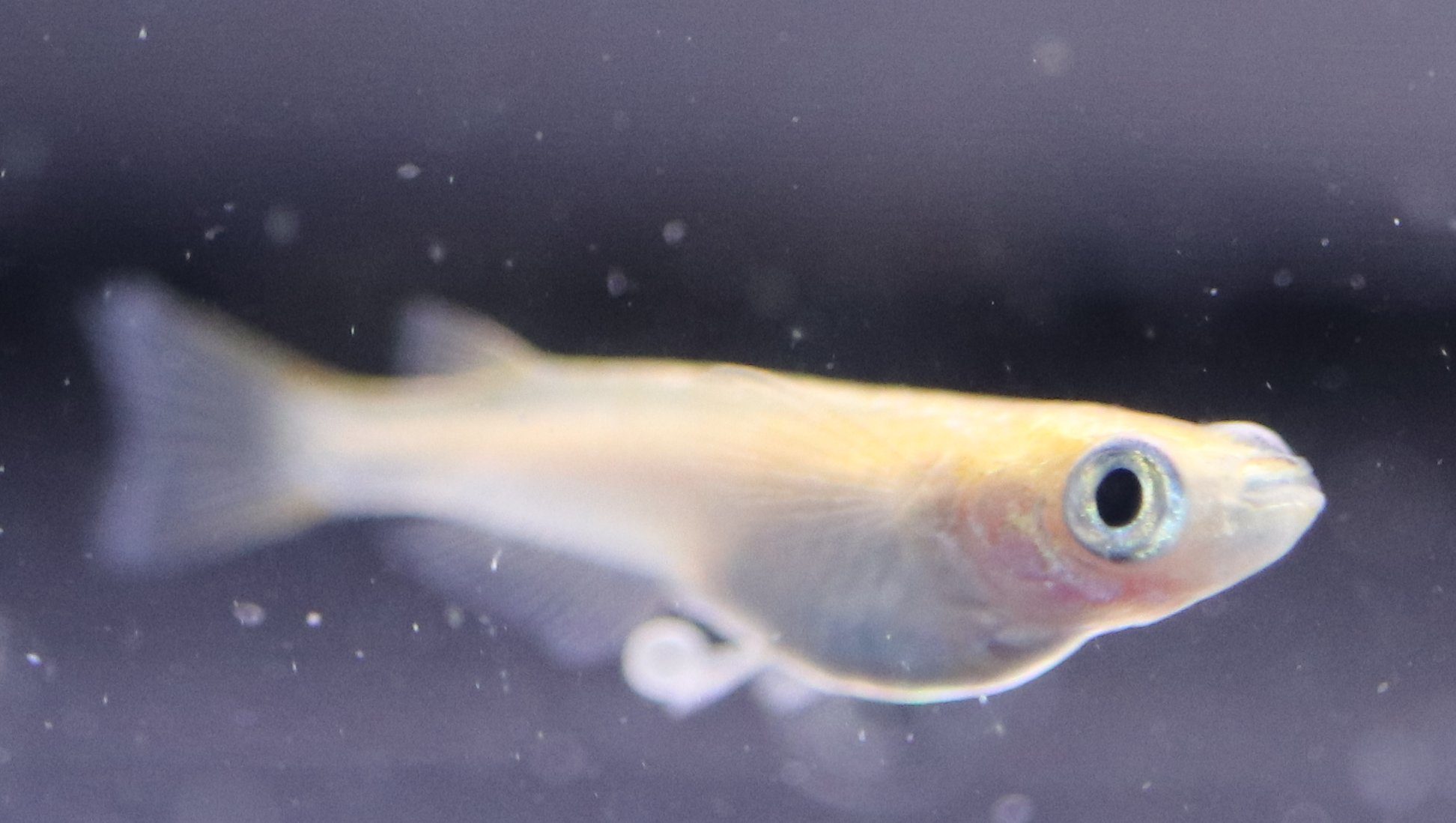
A fish where Japan has particular strength. It is fairly distant from zebrafish, so the two species complement each other: some experiments work in one and not the other. Also the cutest-looking, in our biased view.
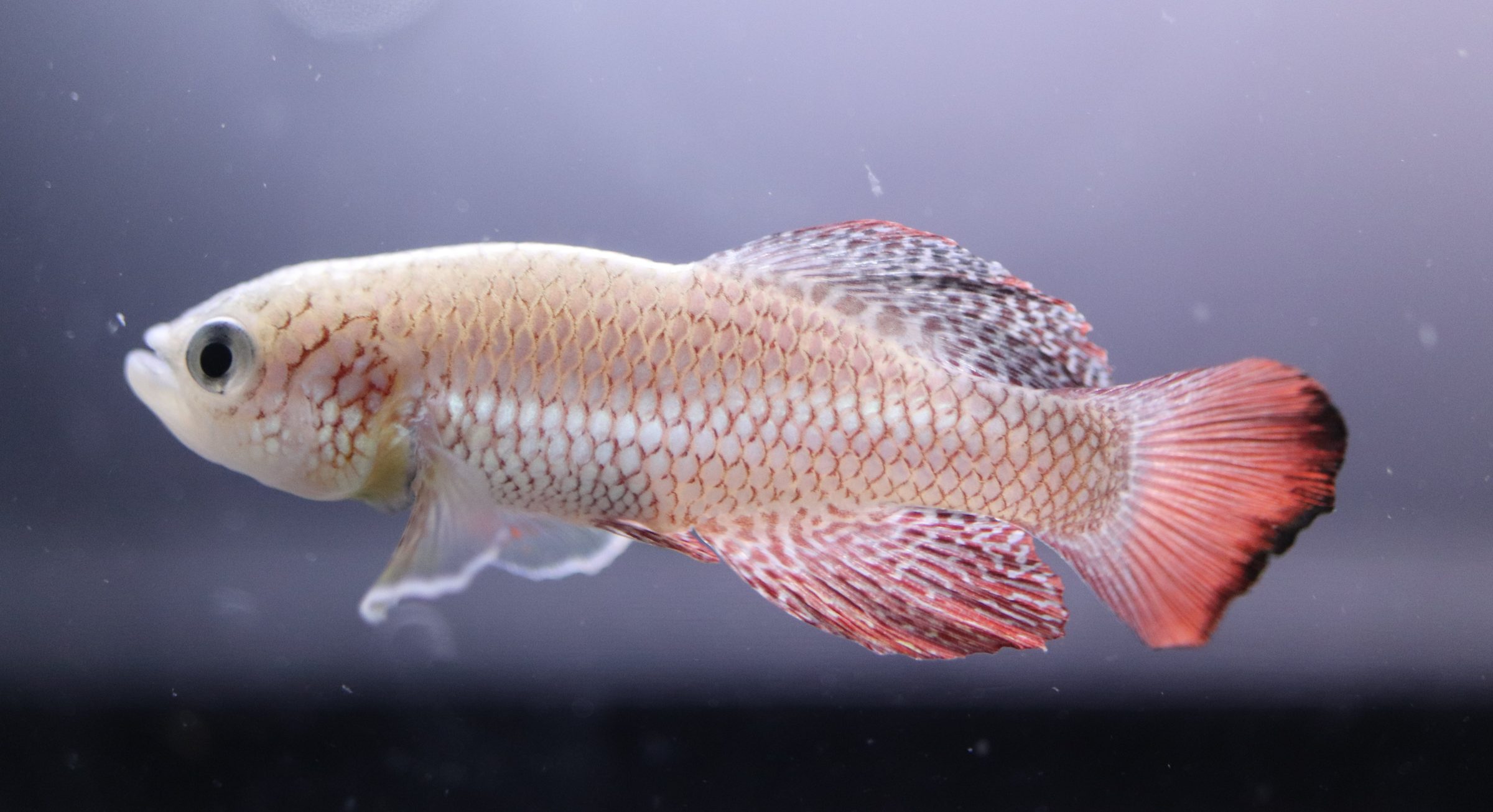
Turquoise killifish
It has a short life history adapted to pools that disappear in the dry season, and it is exceptionally short-lived among vertebrates that can be kept in the laboratory. Rapid aging makes it a strong model for aging and age-related disease. Males are colorful and aggressively intimidating.
Giant danio
We started keeping them for a particular reason, but they are not easy to use experimentally. Picture a large zebrafish. Using a different fish is a good way to remember how unusually good zebrafish are as a laboratory animal.