Summary of previous works

1. アフリカメダカは加齢のみでパーキンソン病の表現型を呈する. African killifish develop Parkinson's disease phenotype only with aging.

私たちはこれまで主にメダカやゼブラフィッシュを用いて薬物の添加や遺伝子改変により様々なパーキンソン病モデルを作製してきました。それらはパーキンソン病の病態の理解に役立ってきましたが、患者さんの大部分を占める孤発性パーキンソン病の病態を模すモデル動物の作製はやはり困難でした。

We have created various models of Parkinson's disease by adding drugs or genetic modification, mainly using medaka and zebrafish. Although these models have been useful for understanding the pathogenesis of Parkinson's disease, it has been difficult to create animal models that mimic the pathogenesis of idiopathic Parkinson's disease, which accounts for the majority of patients.


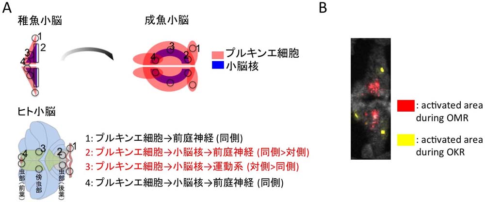
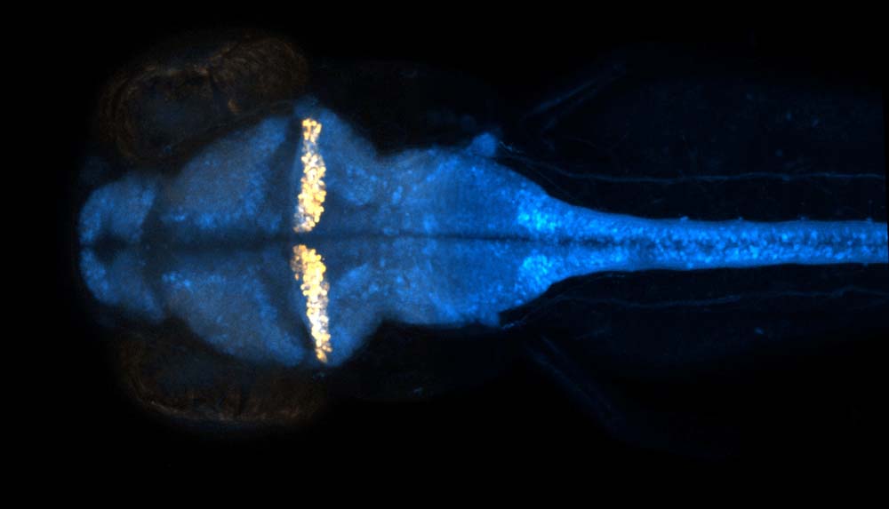
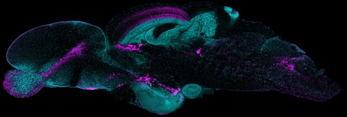
小型魚類の脳のドパミン神経、ノルアドレナリン神経です。左が吻側、右が尾側の脳にです。どれが大脳かわかりますか?

These are dopaminergic and noradrenergic neurons in the brains of small fishes. On the left is the rostral side and on the right is the caudal side of the brain. Can you tell which one is the cerebrum?

ところで魚には様々な利点がありますが、その一つに魚類が多種多様であるということが挙げられます。アフリカのモザンビークには短い雨季の間にだけできる水たまりがあります。その水たまりは乾季には乾燥してしまいます。しかしそこに短い雨季の間だけ魚がいます。彼らはどんなに長生きしたくても、水がなくなれば死んでしまいます。乾燥に強い卵を土の中に埋め、それが雨季に孵化することで種として存続してきました。そのため長生きの形質が選択されず、脊椎動物の中で最も(あるいは2番目に)短命です。

Fish, by the way, have many advantages, one of which is the great variety of fish species. In Mozambique, Africa, there are puddles of water that form only during the short rainy season. These puddles dry up during the dry season. But there are fish there only during the short rainy season. No matter how long they want to live, they will die if there is no water. They have been able to survive as a species by burying their drought-resistant eggs in the soil, which hatch during the rainy season. As a result, they have not been selected for long-lived traits and are the shortest (or second shortest) of all vertebrates.


私たちはパーキンソン病などの加齢依存性の疾患は背景に老化があることで発症しやすくなると考えました。そこでこのアフリカメダカを解析したところ、加齢のみでドパミン神経の変性、αシヌクレインの蓄積といったヒトパーキンソン病に類似の表現型を呈することがわかりました (Matsui et al., Cell Rep, 2019)。またこの魚の脳と、人の脳のαシヌクレインの翻訳後修飾を詳細に解析した結果、パーキンソン病におけるαシヌクレインの神経毒性に関係すると考えられるT64リン酸化の存在を報告しました。また、このT64リン酸化はαシヌクレインのoligomer形成に異常をきたし毒性を発揮することを明らかにしています (Matsui et al., Proc Natl Acad Sci USA, 2023)。

We thought that age-dependent diseases, such as Parkinson's disease, would be more likely to develop due to the presence of aging in the background. When we analyzed the African killifish, we found that it exhibited phenotypes similar to human Parkinson's disease, such as degeneration of dopaminergic neurons and accumulation of alpha-synuclein, due to aging alone (Matsuiet al., Cell Rep, 2019). In addition, detailed analysis of post-translational modifications of α-synuclein in fish brain and human brain revealed the presence of T64 phosphorylation, which is thought to be related to the neurotoxicity of α-synuclein in Parkinson's disease. We have also shown that this T64 phosphorylation causes abnormal oligomer formation of α-synuclein, resulting in toxicity (Matsui et al., Proc Natl Acad Sci USA, 2023).

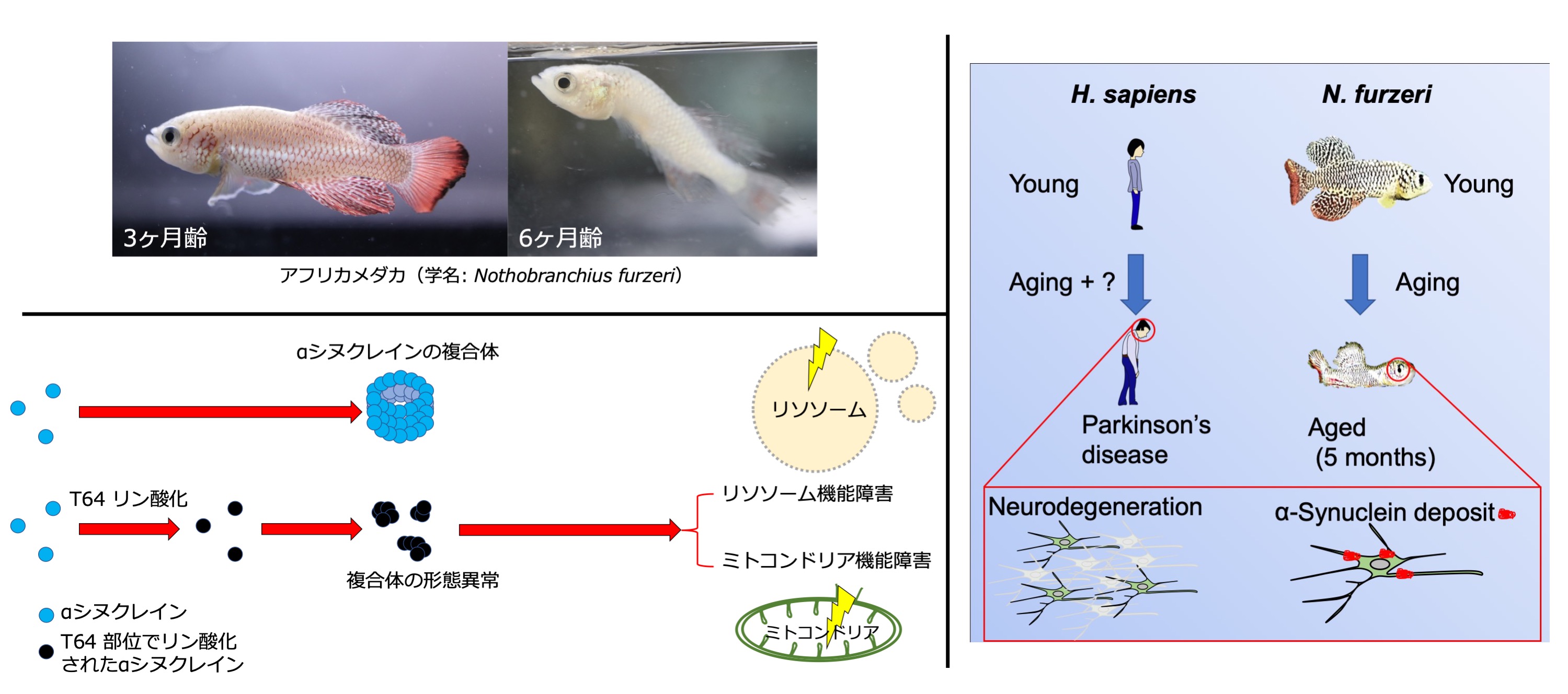
アフリカメダカは5カ月程度の加齢とともに、ドパミン神経の変性やαシヌクレインの蓄積といったヒトのパーキンソン病に類似した病理変化をきたします。 
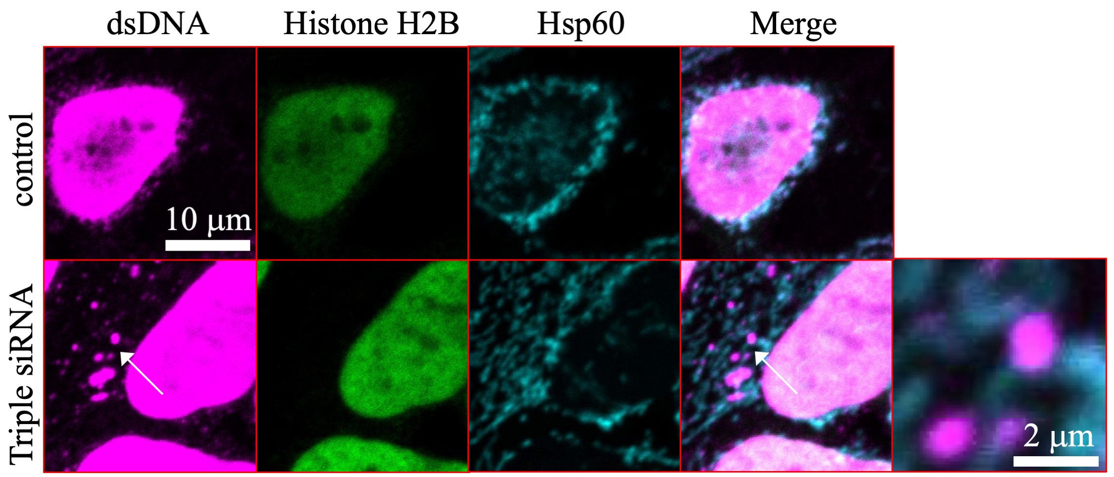
αシヌクレインがT64部位でリン酸化をされることにより、αシヌクレインのT64リン酸化が異常な複合体の形成につながり、結果リソソーム機能障害やミトコンドリア機能障害、ひいては細胞毒性や神経細胞死につながると考えられる。
African killifish develop pathological changes similar to Parkinson's disease in humans, such as degeneration of dopaminergic neurons and accumulation of alpha-synuclein, with aging of about 5 months. The T64 phosphorylation of α-synuclein can lead to the formation of abnormal oligomer complexes, resulting in lysosomal dysfunction, mitochondrial dysfunction, and ultimately cytotoxicity and neuronal cell death.

2. 異所性のミトコンドリアDNAが惹起する神経変性. Neurodegeneration induced by ectopic mitochondrial DNA.

パーキンソン病を模すような処置をした培養細胞やゼブラフィッシュではミトコンドリアのDNAがミトコンドリア外に漏出します。
In cultured cells and zebrafish treated to mimic Parkinson's disease, mitochondrial DNA leaks out of the mitochondria.


これまで私達はパーキンソン病に関する病態解析や新規モデルを多数報告してきました(Nyuzuki et al., IBRO Rep, 2020; Matsui et al., PLOS ONE, 2017, Uemura et al., PLOS Genet 2015; Matsuiet al., FEBS Lett, 2013; Matsuiet al., Hum Mol Genet, 2013; Flinn et al., Ann Neurol, 2013; Matsuiet al., J Neurochem, 2010a; Matsuiet al., J Neurochem, 2010b; Matsui et al., Neurosci Res, 2010; Matsui et al., Neurosci Res, 2009等)。ここでは最新のものについて記載します。

We have reported many pathological analyses and novel models for Parkinson's disease(Nyuzukiet al., IBRO Rep, 2020; Matsuiet al., PLOS ONE, 2017, Uemura et al., PLOS Genet2015; Matsuiet al., FEBS Lett, 2013; Matsuiet al., Hum Mol Genet, 2013; Flinn et al., Ann Neurol, 2013; Matsuiet al., J Neurochem, 2010a; Matsui et al., J Neurochem, 2010b; Matsuiet al., Neurosci Res, 2010; Matsuiet al., Neurosci Res, 2009, etc.). The latest version is described here.

パーキンソン病は運動障害やそれ以外の多彩な症状を呈する神経難病の1つであり、未だにその病態には不明な点が多く残されています。パーキンソン病の病態にミトコンドリア機能障害やリソソーム機能障害が関わっていることは以前より示唆されてきましたが、その詳細なメカニズムはわかっていませんでした。

Parkinson's disease is one of the most common neurological intractable diseases with a wide variety of symptoms, including movement disorders, and many aspects of its pathogenesis remain unknown. It has been suggested that mitochondrial dysfunction and lysosomal dysfunction are involved in the pathogenesis of Parkinson's disease, but the detailed mechanisms have not yet been elucidated.

本研究では、リソソーム中のDNase IIなどによる分解から逃れたミトコンドリア由来の細胞質DNAが、パーキンソン病を模す培養細胞およびゼブラフィッシュにおいて細胞毒性および神経変性を誘導することを報告しました。培養細胞ではパーキンソン病に関連する遺伝子産物であるPINK1、GBA、またはATP13A2の減少は、ミトコンドリア由来の細胞質DNAの増加を引き起こし、I型インターフェロン応答と細胞死を誘導しました。これらの表現型は、DNAを分解するリソソーム内のDNA分解酵素であるDNase IIの過剰発現、またはミトコンドリアDNAのセンサーとして機能するIFI16の減少によって改善しました。パーキンソン病モデルとして用いられるゼブラフィッシュの1つであるgba変異体においても、ヒトDNase IIを過剰発現させることにより、その運動障害とドーパミン作動性神経の変性が改善されました。IFI16およびミトコンドリア由来の細胞質DNAは、パーキンソン病患者の剖検脳の病変部位において蓄積を認めました。

In this study, we report that mitochondria-derived cytoplasmic DNA that escapes degradation by DNase II and other agents in lysosomes induces cytotoxicity and neurodegeneration in cultured cells and zebrafish that mimic Parkinson's disease. In cultured cells, a decrease in the Parkinson's disease-associated proteins PINK1, GBA, or ATP13A2 caused an increase in mitochondria-derived cytoplasmic DNA, inducing a type I interferon response and cell death. These phenotypes were ameliorated by overexpression of DNase II, a DNA-degrading enzyme in lysosomes that degrades DNA, or by a decrease in IFI16, which functions as a sensor of mitochondrial DNA. Overexpression of human DNase II in the gba mutant, a zebrafish model of Parkinson's disease, also ameliorated its motor deficits and dopaminergic degeneration. Accumulation of IFI16 and mitochondria-derived cytoplasmic DNA was observed in lesion sites in autopsied brains of patients with Parkinson's disease.

以上の結果は、ミトコンドリアDNAの細胞質への漏出がパーキンソン病の神経変性の重要な原因となる可能性を示唆しています。細胞質に漏出したミトコンドリアDNAの分解、あるいはそのミトコンドリアDNAセンサーの阻害が、パーキンソン病の治療につながる可能性があります。またパーキンソン病以外の疾患でも同様のメカニズムが存在する可能性があり、引き続き検証を進めます(Matsui et al., Nat Commun, 2021、プレスリリース: https://www.bri.niigata-u.ac.jp/research/result/210521.research_findings.pdf)。

These results suggest that leakage of mitochondrial DNA into the cytoplasm may be an important cause of neurodegeneration in Parkinson's disease. Degradation of mitochondrial DNA leaking into the cytoplasm, or inhibition of its mitochondrial DNA sensor, may lead to the treatment of Parkinson's disease. There is also a possibility that similar mechanisms exist in diseases other than Parkinson's disease, and we will continue to verify this (Matsui et al., Nat Commun, 2021, press: https://www.bri.niigata-u.ac.jp/research/result/210521.research_findings.pdf).


私たちが考えるパーキンソン病の病態仮説です実際にはミトコンドリア機能障害もリソソーム機能障害もどちらも起きていて、異所性ミトコンドリアDNAが処理できなくなる状況が蓄積すると炎症反応や細胞死につながるのではないかと考えています。
This is our hypothesis of the pathogenesis of Parkinson's disease. In fact, we believe that both mitochondrial and lysosomal dysfunction occur, and that the accumulation of conditions that ectopic mitochondrial DNA cannot process may lead to inflammatory reactions and cell death.
3. ゼブラフィッシュの小脳の機能地図. Functional map of the cerebellum in zebrafish.


ゼブラフィッシュの小脳の解剖学的な神経連絡、行動に伴った神経活動、および神経がつかさどる機能、それらを解析しゼブラフィッシュ小脳の機能地図の作成を行いました。
We created a functional map of the zebrafish cerebellum by analyzing the anatomical neural connections, neural activity associated with behavior, and the functions of the neurons.


魚の小脳はヒトの小脳のようにしわや境目がなく、一見すると均一に見えます。魚の小脳の神経回路はヒトのそれとわずかな違いはあるものの、かなりの部分で類似しています。ここではゼブラフィッシュの小脳の遠心路をいくつかの方法で明らかにしました。さらに遊泳時や眼振時に特異的な神経活動をカルシウムイメージングで描出し、解剖学的な地図と神経活動の地図が対応することを示しました。光遺伝学を用いてそれらの部位の神経活動に介入すると、対応する行動の異常を来しました。(Matsui et al., Proc Natl Acad Sci USA, 2014)。

The fish cerebellum does not have wrinkles or borders like the human cerebellum, and at first glance appears to be uniform. The neural circuitry of the fish cerebellum is quite similar to that of humans, although there are slight differences. Here, the efferent pathways of the zebrafish cerebellum have been clarified in several ways. We also used calcium imaging to delineate specific neural activity during swimming and nystagmus, and showed that the anatomical map and the map of neural activity correspond. When we intervened in the neural activity of these regions using optogenetics, the corresponding behavioral abnormalities occurred. (Matsuiet al., Proc Natl Acad Sci USA, 2014).


ゼブラフィッシュの全中枢神経(青)と小脳プルキンエ細胞(金)を異なる蛍光タンパクでラベルしたTransgenic系統です。
Zebrafish whole central nervous system (blue) and cerebellar Purkinje cells (gold) are labeled with different fluorescent proteins.


これらのことから魚の前庭小脳と脊髄小脳の機能地図を描くことに成功しましたが、それ以外にも未知の領域が存在します。ヒトにおいても前庭小脳と脊髄小脳は機能解析が進んできましたが、社会性などの高次機能に関する小脳の役割はまだまだ未解明の部分が多くあります。魚やヒトの小脳、特に大脳と関わる部分は、いったい何をしているのでしょうか?

We have succeeded in drawing a functional map of the vestibular cerebellum and spino-cerebellum in fish, but there are still other unknown areas. In humans, the vestibular cerebellum and spino-cerebellum have been analyzed, but the role of the cerebellum in higher functions such as sociality is still largely unknown. What is the function of the cerebellum in fish and humans, especially in relation to the cerebrum?